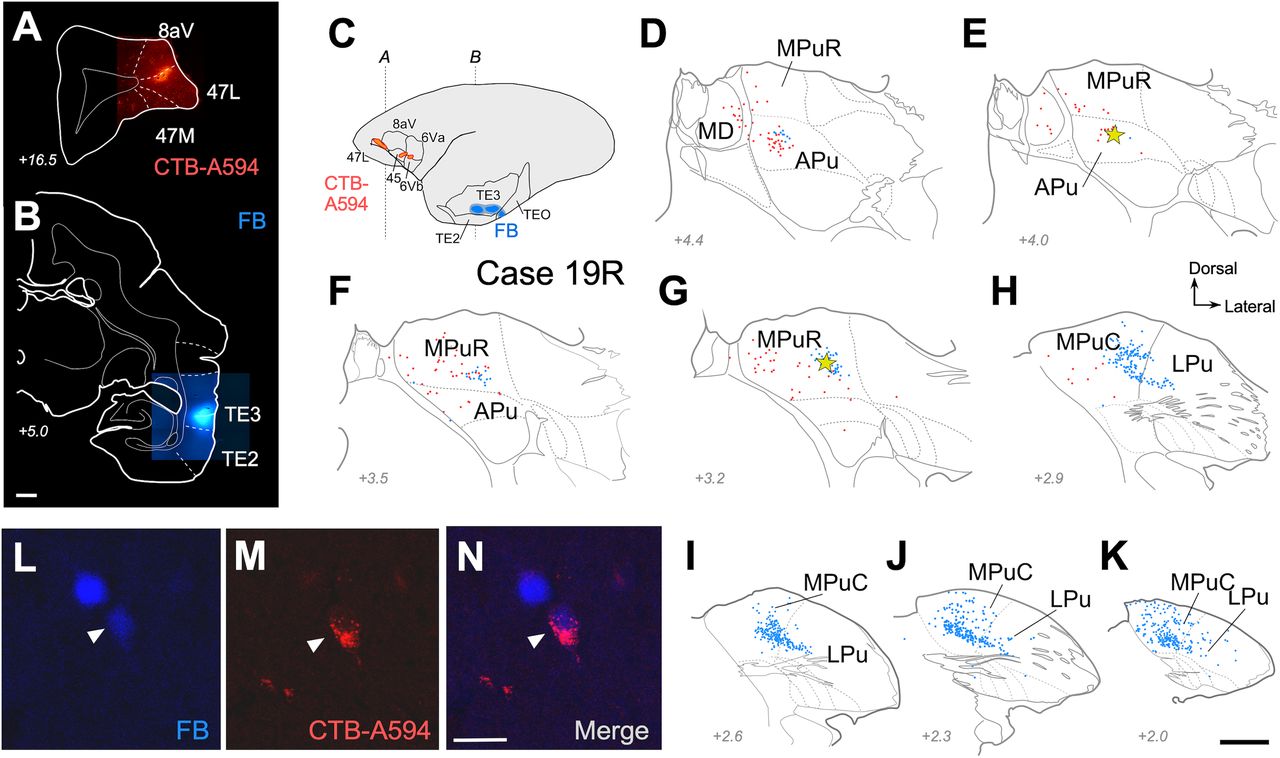
Double-retrograde labeling reveals pulvinar neurons whose axons simultaneously reach the ventrolateral prefrontal and inferior temporal cortices. A, B, Representative coronal section images taken at the center of the tracer deposits in Case 19L. In this experiment, two CTB-Alexa Fluor 594 deposits were made in the lateral prefrontal cortex, involving 47 , 8aV, 6Vb, 6Va, and 45 (A). In the same hemisphere, three contiguous FB deposits involved areas TE3, TE2, and TEO (B). C, Diagram representing the extent of the CTB-Alexa Fluor 594 (red) and FB (blue) deposits in the cerebral hemisphere of Case 19L. D–K, Coronal section diagrams showing the position of labeled cells in the posterior thalamus. Each dot represents a labeled cell body. FB-labeled neurons are drawn in blue and CTB-Alexa Fluor–labeled neurons in red. Yellow stars indicate double-labeled neurons. See Figure 1 for abbreviations. Scale bar, 500 µm. L–N, High-magnification detail showing a double-labeled MPuR cell. Scale bars: A, B, 1 mm; D–K, 500 µm.


