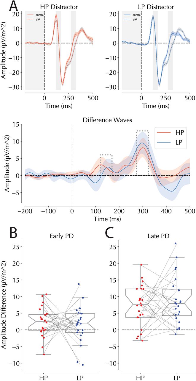
ERPs following distractor and target presentations at the HP and LP locations. A, ERPs following distractor presentation at HP (red) and LP (blue) locations at PO7/8, respectively. Note that trials were restricted to those where the target was on the vertical meridian. The top panels represent condition-specific ERPs for HP (left) and LP (right) trials separating contralateral and ipsilateral sensors (dotted lines represent ipsilateral activity). The hatched shaded bars represent the windows of interest selected for the early (left) and late PD (right) analyses isolated on peaks identified in HP and LP trials separately. The bottom panel shows the difference curve calculated as the contralateral-minus-ipsilateral signal across time. Colored bars represent significant deviations from zero as revealed by a cluster-based permutation test. Significant differences between the two lines would have been denoted by black bars. Shaded areas in the top two panels are within-subject corrected while those in the bottom panel are not. B, Binned lateralized responses in the early PD window for HP and LP trials calculated by subtracting ipsilateral form contralateral waveforms. Dots represent individual participants with lines connecting their scores across the two conditions. C, Binned lateralized responses in the late PD windows of interest. Boxplots in B and C follow the same conventions as Figure 4A.


