Association between Inhibitory–Excitatory Balance and Brain Activity Response during Cognitive Flexibility in Young and Older Individuals
- Geraldine Rodríguez-Nieto1,2,
- David F. Alvarez-Anacona3,
- Dante Mantini1,2,
- Richard A. E. Edden4,
- Georg Oeltzschner4,
- Stefan Sunaert5, and
- Stephan P. Swinnen1,2
- 1Movement Control and Neuroplasticity Research Group, Biomedical Sciences, KU Leuven, Leuven 3001, Belgium
- 2Leuven Brain Institute, KU Leuven, Leuven 3000, Belgium
- 3Facultad de Medicina, Universidad Nacional de Colombia, Bogotá 111321, Colombia
- 4Russell H. Morgan Department of Radiology and Radiological Science, The Johns Hopkins University School of Medicine, Baltimore, Maryland 21287
- 5Department of Imaging and Pathology, Biomedical Sciences, KU Leuven, Leuven 3000, Belgium
-
Author contributions: G.R.-N., D.M., R.A.E.E., G.O., S.S., and S.P.S. designed research; G.R-N. performed research; G.R-N. and D.F.A.-A. analyzed data; G.R-N. and S.S. wrote the paper.
Abstract
Cognitive flexibility represents the capacity to switch among different mental schemes, providing an adaptive advantage to a changing environment. The neural underpinnings of this executive function have been deeply studied in humans through fMRI, showing that the left inferior frontal cortex (IFC) and the left inferior parietal lobule (IPL) are crucial. Here, we investigated the inhibitory–excitatory balance in these regions by means of γ-aminobutyric acid (GABA+) and glutamate + glutamine (Glx), measured with magnetic resonance spectroscopy, during a cognitive flexibility task and its relationship with the performance level and the local task-induced blood oxygenation level-dependent (BOLD) response in 40 young (18–35 years; 26 female) and 40 older (18–35 years; 21 female) human adults. As the IFC and the IPL are richly connected regions, we also examined whole-brain effects associated with their local metabolic activity. Results did not show absolute metabolic modulations associated with flexibility performance, but the performance level was related to the direction of metabolic modulation in the IPL with opposite patterns in young and older individuals. The individual inhibitory–excitatory balance modulation showed an inverse relationship with the local BOLD response in the IPL. Finally, the modulation of inhibitory–excitatory balance in IPL was related to whole-brain effects only in older individuals. These findings show disparities in the metabolic mechanisms underlying cognitive flexibility in young and older adults and their association with the performance level and BOLD response. Such metabolic differences are likely to play a role in executive functioning during aging and specifically in cognitive flexibility.
Significance Statement
Cognitive flexibility provides an advantage in adapting to changing environments. We investigated the inhibitory–excitatory balance (GABA+/Glx) modulation in the frontal and parietal cortices during cognitive flexibility in young and older individuals through magnetic resonance spectroscopy. An increase in the excitatory tone during cognitive performance related to a better execution in younger adults. Interestingly, it was an increase in the inhibitory tone that was associated with a better performance in older adults. Furthermore, we revealed that an increased inhibitory tone in older adults related to a decreased oxygen consumption in remote brain areas (BOLD–fMRI). This may suggest that GABA modulation facilitates the segregation of neural networks, maximizing brain efficiency and cognitive performance. These findings underscore age-related disparities in the neurometabolic mechanisms underlying cognitive flexibility.
Introduction
Cognitive flexibility, one of the main executive function domains, represents the capacity to switch among different mental schemes, providing a distinct advantage in rapidly changing environments. However, this ability may be compromised during aging, which becomes critical in a highly dynamic world with an increasing proportion of older individuals.
Cognitive flexibility requires the engagement of the middle and inferior frontal gyri, extending to the inferior frontal junction (IFJ), as well as the inferior parietal lobule (IPL; Rodríguez-Nieto et al., 2022). The IFJ is crucial for adjusting behavior according to abstract rules (Derrfuss et al., 2005; Worringer et al., 2019). The IPL is involved in a wide variety of cognitive domains, such as motor imagery, attention shifting, and target discrimination (Capotosto et al., 2013; Kraeutner et al., 2019; Gray et al., 2020). Both regions are functionally connected across a wide variety of higher-order cognitive tasks (Cieslik et al., 2016; Camilleri et al., 2018).
The left IFJ shows changes in executive function-related brain activations as an effect of aging, and hypometabolism in this region is associated with executive deficits in different neurodegenerative disorders (Schroeter et al., 2012; Heckner et al., 2021). Moreover, brain damage and temporal disruption by transcranial magnetic stimulation of the left IFJ support its crucial role in executive functioning (Muhle-Karbe et al., 2014; Kubler et al., 2019; Schroeter et al., 2020).
Little is known about the neurometabolic dynamics during cognitive flexibility. GABA and glutamate are fundamental in the regulation of neural activity, either by decreasing or increasing neuronal excitability, respectively. Magnetic resonance spectroscopy (MRS) measures these metabolites in the in vivo human brain, providing an estimate of tonic levels (i.e., average quantification across time in the order of minutes). With this technique, it was observed that a relationship between higher levels of baseline GABA+ (GABA plus macromolecules) and Glx (glutamate + glutamine) in the anterior cingulate cortex (ACC) related to faster responses or better performance during flexible behavior (Marenco et al., 2018; Stock et al., 2023). On the other hand, a review proposed that lower baseline GABA levels can facilitate flexible behaviors (Li et al., 2022). Importantly, the neurometabolic task-related changes (as measured with functional MRS) have not been studied in the context of cognitive flexibility yet. It is reasonable to assume that cognitive flexibility involves an interplay of excitatory and inhibitory mechanisms. Whereas an inhibition reduction may be necessary to activate different local and global networks, a certain level of GABA may be necessary to suppress irrelevant information during shifting. Moreover, due to their simultaneous influence on the brain, the GABA/glutamate balance may ultimately determine the activation of specific regions in the brain.
Older adults exhibit lower levels of GABA and glutamate (Hermans et al., 2018; Cleeland et al., 2019), although this seems region dependent for glutamate (Rodríguez-Nieto et al., 2023). Older adults also exhibit decreases in their executive performance level (Seer et al., 2021), and this has been shown to be related to baseline GABA levels (Hermans et al., 2018; Heise et al., 2022). Therefore, metabolic modulation occurring during executive task performance may be age-dependent.
We hypothesize that the MRS-assessed GABA+/Glx balance plays a role in cognitive flexibility and that tonic GABA+/Glx is reduced during cognitive flexibility performance in the regions showing a task-induced blood oxygenation level-dependent (BOLD) response, i.e., inferior frontal cortex (IFC) and IPL. We also investigate whether this modulation is related to individual differences in cognitive flexibility performance. Furthermore, we examine whether the metabolic modulation in the IFC and IPL corresponds to the BOLD response associated with flexible behavior. As IFC and IPL are association areas known to be richly connected with remote brain regions (Yeo et al., 2015), we also investigate whether local metabolic modulations in IFC and IPL are associated with the neural activation in remote brain regions. Importantly, we address these objectives in young and older adults, to identify age-related differences in the metabolic modulation associated with cognitive flexibility.
Materials and Methods
Participants
Forty young (YA; mean age, 23.38 years; SD, 3.81; range, 18–35 years old; 26 females) and forty older (OA; mean ± SD age, 69.52 years; SD, 5.42; range, 60–85 years old; 21 females) healthy participants were recruited for this study. All participants reported absence of neurological or psychiatric disorders. In addition, they were subjected to the Montreal Cognitive Assessment to exclude participants with mild cognitive impairment (YA, mean score, 27.4; range, 24–30; OA, mean score, 27.1; range, 22–30). One older participant scored below the cutoff point (score, 22; cutoff, 24; Islam et al., 2023) and was excluded for that reason. The handedness of participants was assessed with the Edinburgh Handedness Questionnaire. The handedness proportion of participants was representative of that reported for the general population (Willems et al., 2014): 37 young adults and 37 older adults were right-handed; three younger adults and two older adults were left-handed, and one older adult was ambidextrous. The experiment was approved by the local ethical committee of KU Leuven (Protocol Number s61577), and all participants gave written informed consent.
Experimental design
Prior to the MRI session, participants performed 16 practice trials of the rule-switching task used to assess cognitive flexibility. Once positioned in the MRI scanner, they performed the task while the fMRI data were being collected. Afterward, the MRS protocol was applied, consisting of (1) two MRS scans at the baseline (resting-state MRS) with the voxels centered at the IFC and IPL, (2) a short T1-weighted scan to minimize the effect of intermediate participant movement on voxel location by using it for the coregistration of images collected during the MRS scans in (3), and (3) two MRS scans with the voxels centered at the IFC and IPL while participants performed the task (task-related functional MRS; Fig. 1a). The order of the two MRS scans in (1) and (3) was counterbalanced across participants.

A, MRI session protocol. rMRS, resting-state MRS; fMRS, MRS during cognitive flexibility performance. B, Cognitive flexibility task design.
The selection and positioning of the two MRS voxels were based on our extensive meta-analysis results for rule-switching paradigms that showed a consistent activation in the IFC and the IPL (Rodríguez-Nieto et al., 2022). Furthermore, these two regions were preferred over other relevant nodes of cognitive flexibility (such as the insula, the striatum, or ACC) due to their higher specificity for higher-order cognitive functioning and to avoid the potential inclusion of adjacent regions containing cerebrospinal fluid.
Behavioral task
In the rule-switching flexibility task (adapted from Sekutowicz et al., 2016; launched in PsychoPy; Peirce, 2007), participants were asked to respond through a button box whether the number shown on the screen was lower or higher than five (left or right button) when the number was preceded (cue) and simultaneously surrounded by a circle and to indicate whether it was odd or even (left or right button) if the number was preceded (cue) and simultaneously surrounded by a square (Fig. 1b). In half of the trials, the rule was the same as in the previous trial (repeat trial) and in the other half the rule switched (switch trial) based on a pseudorandomized sequence. The task in the fMRI consisted of 94 trials [cue alone, 300 ms; cue + stimulus, 1,700 ms; intertrial interval (ITI), 3 or 4 s] and of 132 trials in each of the two MRS blocks (cue alone, 400 ms; cue + stimulus, 2.2 s; and ITI, 2, 3, or 4 s).
Imaging data acquisition
All MRI scans were acquired using a Philips Achieva 3 T scanner with a 32-channel head coil. For structural neuroimaging, a high-resolution three-dimensional T1–weighted image was collected (MPRAGE, time repetition/time echo, 9.6/4.6 ms; 0.98 × 0.98 × 1.2 mm3 voxel size; field of view, 192 × 250 × 250; 160 coronal slices; scan time, ∼7 min). Before the task-related MRS, a short T1-weighted anatomical image was acquired to verify a correct alignment for the MRS voxels.
To acquire functional scans (fMRI), an ascending gradient echo-planar imaging pulse sequence for T2*-weighted images [repetition time (TR), 1,000 ms; echo time (TE), 33 ms; flip angle, 70°; 42 transverse slices; interslice gap, 0.5 mm; voxel size, 2.15 × 2.14 × 3 mm3; field of view, 240 × 240 × 146.5 mm3] was used.
For MRS, a Hadamard Encoding and Reconstruction of MEGA-Edited Spectroscopy (TR, 2,000 ms; TE, 80 ms; 320 water-suppressed signal averages, 5 kHz spectral width; 4,096 datapoints; 16 unsuppressed scan averages) sequence was used (Saleh et al., 2016).
Four MRS blocks (two conditions × two voxels) were conducted to target GABA+ and Glx (glutamate + glutamine) levels in the left IFC (4 × 2.5 × 2.5 cm3 voxel) and in the left IPL (3 cm3 voxel). The procedure to locate the voxel for the IFC was as follows: (1) center the voxel at the first axial slice above the ventricles at the height of one-third of the sagittal midline and at the width center of the left hemisphere; (2) from the sagittal view, locate the center length of the voxel parallel to the inferior frontal sulcus and locate the three-fourth length over the IFJ; and (3) from the coronal view, locate the top of the voxel parallel to the cortical surface. When needed, this procedure was adapted to avoid covering the ventricles or the subarachnoid space. To locate the IPL voxel we (1) located the voxel four slices above the last axial view of the ventricles and the first view after the ventricles in the coronal plane, (2) placed one side parallel to the occipitoparietal sulcus, (3) moved the voxel toward the axial midline of the left hemisphere, and (4), in the sagittal plane, located it parallel to the inferior parietal sulcus and inspected that this was mostly covered.
Statistical analyses
For the rule-switching task, we calculated the flexibility index by subtracting the number of hits in repeat trials from the number of hits from the switch trials. A higher score indicated a better cognitive flexibility performance. One old participant was excluded due to a low flexibility score (<3 SD). A mixed repeated-measure general linear model (GLM) was conducted to examine intrasubject differences across the three task stages (during fMRI and the two blocks of MRS) and to examine (intersubject) differences between young and older participants. Regression analyses were performed to test the relationship between cognitive flexibility performance and GABA+/Glx modulation, as well as the relationship between this modulation and local BOLD response.
fMRI data analysis
fMRI data were analyzed with FMRIB Software Library v.6.05. First, the Brain Extraction Tool was used on the T1-weighted anatomical images to extract the brain from the dura and skull. Next, the fMRI data were preprocessed: they were high-pass filtered with cutoff time constant of 90 s, motion-corrected with MCFLIRT, and spatially smoothed at 8 mm full-width half-maximum. Functional images were coregistered to their corresponding T1-weighted anatomical image using a linear transformation (12 DOF) and visually inspected. The resulting image was normalized (linear registration, 12 DOF) to the MNI152 template. fMRI data of six young participants were excluded due to technical difficulties leading to nonusable data (4) and excessive head movement (2; cutoff, 1.5 mm for absolute and 0.65 for relative motion; excluded values, 1.6 and 1.7 mm absolute motion).
For each individual, we applied a fixed-effect GLM in which repeat trials, switch trials, and ITIs were included as regressors along with their first temporal derivative. The statistical maps were rendered using a z-threshold of 2.3 and a (corrected) cluster significance threshold of p = 0.05. The motion parameters were included as regressors in the first-level GLMs. Additionally, we inspected the plots of estimated rotations, translations, and mean displacements for each participant. The absolute motion range (mm) was as follows: young, 0.09–0.64 (x̄ = 0.24; SD, 0.13); old, 0.14–0.69 (x̄ = 0.33; SD, 0.24); and cutoff, 1.5. The relative motion range (mm) was as follows: young, 0.05–0.2 (x̄ = 0.07; SD, 0.03); old, 0.09–0.37 (x̄ = 0.14; SD, 0.05); and cutoff, 0.65.
A random-effect model was used to extract each group's mean BOLD activity in the Switch > Repeat contrast. Next, group comparisons OA > YA and YA > OA were performed for this contrast (maps were cluster-corrected, threshold p = 0.05, and rendered using a z value of 2.7).
The beta values from the Switch > Repeat contrast were extracted for each participant from a mask delineated from the sum of individual IFJ and IPL voxels, in order to examine the relationship between BOLD and metabolic activity locally. In further analyses, we included the demeaned levels of GABA+/Glx (baseline and modulation) from each voxel as covariate in the whole-brain analyses (Switch > Repeat) to address global BOLD correlates of local metabolic activity at IPL and IFC. For this analysis, group comparisons (OA > YA and YA > OA) were also performed (maps were cluster-corrected, threshold p = 0.05, and rendered using a z value of 2.7).
MRS data analysis
Data were preprocessed and quantified using Gannet v. 3.3.0 (Edden et al., 2014), including line broadening (3 Hz), eddy current correction, and spectral registration (Mikkelsen et al., 2020). All preprocessed data were visually inspected to detect artifacts and to assess the spectrum appearance. Two datapoints from GABA+ (which included the spectra of GABA and macromolecules) in the IFC task block were excluded for presenting a fitting error (defined as the standard deviation of the model residual divided by the amplitude of the fitted signal) above 3 SD.
The voxel masks for each analysis were coregistered to their respective anatomical image to determine gray matter, white matter, and cerebrospinal fluid fractions using SPM12 (Friston et al., 1994). The metabolite levels were tissue-corrected according to the α-correction method (Harris et al., 2015). Figure 2 shows the overlap of the voxels from all participants positioned for IFC and IPL, as well as the average spectra for each condition (baseline and condition) and by age group, highlighting consistent voxel placement and excellent spectral quality.

Voxel overlap positioning from all participants and average spectra from young (yellow) and older (purple) participants during baseline (top panel) and task performance (bottom panel).
In order to measure the changes in inhibitory–excitatory balance as the result of cognitive flexibility performance, a GABA+/Glx modulation index (one per region) was estimated by subtracting the GABA+/Glx ratio obtained during the resting-state blocks from the GABA+/Glx ratio obtained during task performance. A positive modulation index indicates an increase in GABA+/Glx during task performance as compared with the baseline, whereas a negative index indicates a decrease.
Whereas the BOLD response and measurements of metabolite levels by MRS (as well as derived indices) are part of metabolic activity, we will refer specifically to the indices derived from the MRS measurements (GABA+ and Glx) when we make use of this term.
A mixed repeated-measure GLM was conducted to examine differences in the GABA+/Glx ratio between baseline and during the performance of the task (intraindividual factor) and between young and older adults (interindividual factor).
Results
Behavior: cognitive flexibility
A mixed repeated-measure GLM revealed group (F(1,71) = 10.71; p = 0.002; η2 = 0.14), block (F(1,71) = 80.55; p < 0.001; η2 = 0.56), and group × block (F(1,71) = 9.87; p = 0.002; η2 = 0.13) effects for cognitive flexibility performance. In particular, young adults performed better than older adults during the first block (fMRI; t(70) = −3.33; p ≤ 0.001; d = 0.79; YA, mean, −7.17; SD, 8.61; OA, mean, −14.31; SD, 9.22) and the second block (MRS1; t(70) = −3.13; p = 0.001; d = 0.74; YA, mean, 0.14; SD, 0.62; OA, mean, −0.32 ± 0.62) but not during the third one (MRS2; t(70) = −0.26; p = 0.39; d = 0.07; YA, mean, −0.58; SD, 0.68; OA, mean, −0.53; SD, 0.84). Both groups showed a higher number of errors in switch trials as compared with control (repeat) trials during the first block (fMRI block mean score, −11.15), but during the next blocks, both groups had a considerably lower rate of errors during switching trials, comparable to the low rate or errors in control trials (MRS1 block mean score, −0.09; MRS2 block mean score, −0.56). Therefore, to exclude time-related effects from cognitive flexibility performance (i.e., learning), we considered the behavioral index from the first block (fMRI) in further analyses, which reflects better the variability in the default cognitive flexibility of participants (Fig. 3).

Behavioral performance in the cognitive flexibility task in young (yellow) and older (purple) adults. Younger individuals performed better than older individuals (F = 13.42; p < 0.001; η2 = 0.16), and individuals showed a better performance during repeat trials as compared with switch trials (F = 99.29; p < 0.001; η2 = 0.59), with this difference being more accentuated in older individuals (F = 10.93; p = 0.002; η2 = 0.14).
GABA+/Glx modulation during cognitive flexibility and age-related effects
A mixed GLM model (repeated measures) was conducted to address differences in GABA+/Glx according to group (intersubject variable), condition (intrasubject variable), and their interaction in IFC and IPL with the order (measured during first or second block) as covariate.
There was neither a significant main effect of group, condition, nor their interaction, on GABA+/Glx levels in IFC (group, F(1,76) = 0.02; p = 0.89; η2 < 0.001; condition, F(1,76) = 1.02; p = 0.32; η2 < 0.001; interaction, F(1,76) = 1.9; p = 0.16; η2 = 0.02) and neither in IPL (group, F(1,77) = 0.31; p = 0.58; η2 = 003; condition, F(1,77) = 0.21; p = 0.64; η2 < 0.001; interaction, F(1,77) = 0.16; p = 0.68; η2 = 0.02; Fig. 4). Interestingly, previous reports (e.g., Gao et al., 2013; Rowland et al., 2016; Hermans et al., 2018; Rodríguez-Nieto et al., 2022) showed that GABA+ and Glx levels (when analyzed separately) are lower in older individuals.

A, GABA+/Glx levels in IFC (top) and in IPL (bottom) for young (yellow) and older (purple) participants during baseline and task performance. The differences according to the age group, condition, or their interaction were not significant (IFC, group, F = 0.02; p = 0.89; η2 < 0.001; condition, F = 1.02; p = 0.32; η2 < 0.001; interaction, F = 1.9; p = 0.16; η2 < 0.02; IPL, group, F = 0.31; p = 0.58; η2 = 003; condition, F = 0.21; p = 0.64; η2 < 0.001; interaction, F = 0.16; p = 0.68; η2 = 0.02). B, Individual modulation (change from the baseline to task performance) in IFC (top) and IPL (bottom) for younger (left) and older (right) adults. Horizontal black lines indicate the mean value.
Relationship between task-related GABA+/Glx modulation and cognitive flexibility level
According to the regression analysis, the modulation of GABA+/Glx in IPL and its interaction with age were significant predictors of cognitive flexibility performance, while the same variables in IFC lacked predictive value within the same model (R2 = 0.13; F(5,65) = 4.84; p = 0.01; Table 1; Fig. 5). To examine the age effect, we ran a regression analysis in the two age samples separately to predict cognitive flexibility level with modulation of GABA+/Glx in IPL as independent variable (Table 1; Fig. 5). Whereas a decrease of GABA+/Glx in IPL predicted a better performance in young participants (β = −0.36; p = 0.05), this pattern was inversed in older individuals (β = 0.37; p = 0.02; Table 1; Fig. 5).

Regression of flexibility performance from GABA+/Glx modulation (R2 = 0.13; F = 4.84; p = 0.01; top panel) and GABA+ (bottom panel) for younger (yellow; R2 = 0.09; F = 2.87; p = 0.11) and older (purple; R2 = 0.14; F = 6.03; p = 0.02) participants. Higher ΔGABA+/Glx indicates a higher increase in the inhibitory tone, and the higher the flexibility score, the better the performance.
Prediction of cognitive flexibility performance level by modulation of GABA+/Glx (regression analysis)
To test whether the significant effects between behavior and the changes of GABA+/Glx balance in IPL could have been driven by the change in GABA+ or Glx separately, we ran regression analyses with these variables as predictors in the same model, which were not included in the previous model to avoid multicollinearity. We included ΔGABA+, ΔGlx, and their respective interaction with age in a regression model for the full sample. Only ΔGABA+ and its interaction with age resulted in modulation in older adults (increase in GABA+ significant predictors; R2 = 0.09; F(2,70) = 3.32; p = 0.04; Fig. 5; Table 2). Separate analyses in each age group revealed that in the older adults, an increase of GABA+ predicted a better performance (R2 = 0.14; F(1,37) = 6.03; p = 0.02; Table 2; Fig. 5), but in young individuals, ΔGABA+ did not have a significant predictive value (R2 = 0.09; F(1,33) = 2.87; p = 0.11; Table 2; Fig. 5).
Prediction of the cognitive flexibility performance level by modulation of GABA+ and Glx (regression analysis)
Task-related BOLD response during cognitive flexibility
During switching trials (Switch > Repeat), young and older adults showed activation in the bilateral inferior and superior parietal lobules: presupplementary area and ACC. Whereas the older participants showed a bilateral activation in the IFJ, young participants only showed this activation in the left hemisphere, although this difference was not significant. In general, the overall BOLD response in young participants was less diffuse and more circumscribed to the left hemisphere. The statistical analysis to directly compare the two age groups showed that older adults had a stronger BOLD response in the bilateral IPL and occipital cortex (z = 2.7; cluster correction threshold p = 0.05; Fig. 6).

BOLD response in both age groups and for the Old > Young comparison (z = 2.7; cluster significance threshold p < 0.05). There were no significant differences for the Young > Old comparison.
Relationship between task-related GABA+/Glx modulation and local BOLD responses
GABA+/Glx modulation in the IFC and its interaction with age did not predict the local BOLD response (IFC voxel; R2 = 0.03; F(2,69) = 1.08; p = 0.34; Table 3). However, in the IPL, a decrease in GABA+/Glx predicted a higher local BOLD response (R2 = 0.07; F(2,71) = 5.33; β = −0.26; p = 0.02; Table 3; Fig. 7), while no significant interaction with age was obtained (β = 0.32; p = 0.38). This effect appeared to be specifically driven by the modulation of GABA+ (R2 = 0.05; F(2,71) = 4.46; β = −0.24; p = 0.04; Table 3).

Regression of local BOLD response in IPL (extracted from the voxel in green) from individual modulation in GABA+/Glx (change from the baseline to task; a positive value indicates an increase; R2 = 0.07; F = 5.33; p = 0.02). The whole-brain BOLD response is shown in the figure on the left for young (yellow) and older (purple) adults.
Prediction of local BOLD response from metabolic modulation
Relationship between task-related GABA+/Glx modulation and whole-brain BOLD response
As IFC and IPL are association areas richly connected with distal brain regions, we investigated whether local GABA+/Glx modulation was associated with brain activity from distal regions. In young adults, no relationship between the task-related GABA+/Glx modulation (in either region) with whole-brain BOLD activation was observed. In older participants, no relationship was found between task-related GABA+/Glx modulation in IFC and global BOLD response. However, an increase in GABA+/Glx during the task as compared with the baseline was related to a decreased BOLD activity in the bilateral occipital cortex and bilateral cerebellum (z = 2.7; cluster correction threshold p = 0.05). Follow-up analyses showed that this relationship was partly driven by a modulation in GABA+, indicating that an increase in GABA+ was related to a lower BOLD response; there was an association overlap in the occipital lobes and cerebellum. In addition, GABA+ was inversely related to BOLD activity in the right middle frontal gyrus (z = 2.7; cluster correction threshold p = 0.05; Fig. 8). The group comparison analyses revealed that the different activation patterns observed in young and older adults were not significantly different (z = 2.7; cluster correction threshold p = 0.05).
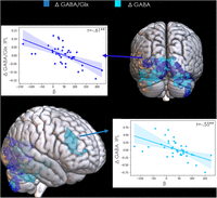
Regions that showed a significant and negative correlation with the modulation of GABA+/Glx and GABA+ in IPL during cognitive flexibility (Switch > Repeat) in older adults. A decrease in the inhibitory tone was related to an enhanced BOLD activation in these regions. Statistic images were generated with a z-threshold of 2.7 and a corrected cluster significance threshold of p = 0.05.
Relationship between baseline GABA+/Glx and whole-brain BOLD response
We conducted complementary analyses to examine whether baseline (resting-state) GABA+/Glx in IFC and IPL had an effect on whole-brain BOLD response. In young participants, baseline GABA+/Glx in IFC positively correlated with the BOLD response in the left insula, the left IFJ, left ventromedial prefrontal cortex, left amygdala, bilateral basal ganglia, cerebellum, thalamus, ACC, and dorsomedial prefrontal cortex. No effects were shown for baseline GABA+/Glx in IFC in older participants. The difference between the activation observed in young adults and the (null) activation in old participants was statistically different showing that young adults had more activation associated with baseline GABA+/Glx from IFC in the bilateral thalamus, right caudate, right putamen, and the ACC extending to the supplementary motor cortex and to the precentral gyrus (Fig. 9). Also, in young participants, baseline GABA+/Glx in IPL positively correlated with BOLD activity in the thalamus but in older adults baseline GABA+/Glx in IPL positively correlated with the BOLD response in the right middle frontal gyrus, the left cerebellum, and the left temporal and occipital cortices. The comparison of the activation from both age groups did not yield significant differences (Fig. 9).
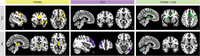
Regions showing a significant correlation with baseline levels of GABA+/Glx in IFC (top) and IPL (bottom) (z = 2.7; cluster significance threshold p < 0.05). No BOLD response correlates were observed for baseline GABA+/Glx in IFC for older adults.
Relationship between whole-brain BOLD-GABA+/Glx–related activation and behavior
We explored whether the whole-brain BOLD-GABA+/Glx–related activation (displayed in Fig. 8) was associated with behavioral performance. In older adults, the whole-brain BOLD response that showed to be associated with a task-induced decrease in GABA+/Glx and GABA+ was significantly associated with behavioral performance (BOLD-ΔGABA+/Glx, r = −0.42; p = 0.008; BOLD-ΔGABA+, r = −0.38; p = 0.02), i.e., older adults who displayed a higher BOLD response (associated with a decrease in inhibitory tone in IPL during the task) tended to have a lower flexibility performance level. Although there were no significant whole-brain BOLD activations associated with ΔGABA+/Glx in younger adults, we conducted the analog correlation from the BOLD response in the same regions with behavior in young adults (as a control analysis), which resulted in a null association (r = 0.14; p = 0.44). The comparison between the correlation values from YA and OA showed that the difference was not statistically significant (z = −1.03; p = 0.15).
As task-related metabolic modulation (ΔGABA+/Glx and ΔGABA+) in IPL was also related to performance in older adults, we performed partial correlations to exclude that the metabolic activity would be driving the relationship between BOLD and behavioral performance. We found that the correlation between whole-brain BOLD-metabolic–related activation and flexibility performance was no longer significant after controlling for task-related metabolic modulation (BOLD-ΔGABA+/Glx, r = −0.27; p = 0.11; BOLD-ΔGABA+, r = −0.31; p = 0.07).
We conducted analog analyses for the relationship between whole-brain BOLD response associated with baseline GABA+/Glx and flexibility performance. No significant correlation was found in young adults (BOLD-GABA+/Glx baseline IFC, r = 0.06; p = 0.75; BOLD-GABA+/Glx baseline IPL, r = 0.05; p = 0.78) nor in the control analyses in older adults (BOLD-GABA+/Glx baseline IFC, r = −0.06; p = 0.73; BOLD-GABA+/Glx baseline IPL, r = −0.03; p = 0.86). In older adults, a higher BOLD-baseline IPL GABA+/Glx associated response related to worse performance (r = −0.38; p = 0.01), which survived after controlling for baseline GABA+/Glx in IPL (r = −0.43; p = 0.009). The control analysis in young participants did not show a significant association (r = −0.14; p = 0.44). The difference between the coefficients from both samples was not significant (z = −1.05; p = 0.14).
Discussion
We aimed to investigate the modulation of inhibitory/excitatory balance (ΔGABA+/Glx) in the IFC and IPL during a cognitive flexibility task as well as the relationship of this modulation with task performance and with local and global BOLD responses in young and older adults. Contrary to what we expected, we did not observe a reduction (nor increment) of GABA+/Glx at the group level. Nonetheless, we found that the task-induced modulation of GABA+/Glx predicts flexibility performance with opposite patterns in young and older adults. Moreover, we found a negative relationship between the task-related GABA+/Glx modulation and local BOLD response in IPL. Finally, we found an effect of task-related GABA+/Glx modulation in the whole-brain BOLD response of older adults. Although no effects were observed in young participants in this regard, their baseline levels of GABA+/Glx were related to whole-brain BOLD effects during cognitive flexibility.
Task-related metabolic modulation and its association with the flexibility performance level
Contrary to what we hypothesized, no overall decrease (nor increase) in GABA+/Glx was observed during cognitive flexibility performance at the group level. Interestingly, whereas young and older adults had similar baseline GABA+/Glx ratios, these groups showed opposite modulation patterns in relation to their performance level.
More specifically, an increase in GABA+/Glx in the IPL was related to worse performance in young adults, while it was related to better performance in older adults. Moreover, this effect was mainly led by a GABA+ modulation in older adults (increase in GABA+ related to a better flexibility performance), and this was not the case in young individuals. Thus, older adults appeared to benefit from an increased inhibition in the same region. Possibly, the upregulation of GABA+ levels in older adults (given that their baseline levels are lower) served to keep it at a necessary minimal threshold level to suppress interference of irrelevant stimuli and optimally perform the task. More research is required to support these assumptions.
Even though we showed that metabolic modulation in the left IPL relates to the performance level in cognitive flexibility, we did not observe absolute metabolic modulation nor correlations between flexibility performance and the metabolic activity in the left IFC. Although these two regions have shown to be consistently active during cognitive flexibility, some studies have pointed to a role of the IFJ and inferior frontal gyrus in abstract switching and a role of IPL (and in particular the inferior parietal sulcus) in the selection and updating of action rules (Philipp et al., 2013; Muhle-Karbe et al., 2014). It may be possible that IFC played a stronger role in an early phase of flexibility performance, when there was a higher executive demand. As the metabolites were measured at a later stage of flexibility performance, where some learning effect was observed, metabolic modulations in IFJ may have been dissipated by then.
BOLD response associated with cognitive flexibility and aging
The BOLD response observed during cognitive flexibility is consistent with previous reports (Worringer et al., 2019; Rodríguez-Nieto et al., 2022). Moreover, as in previous studies, we also observed a more diffuse and extended BOLD response in old as compared with young adults during cognitive flexibility performance (Eich et al., 2016; Kunimi et al., 2016; Kuptsova et al., 2016). The increased bilateralized processing in older adults is in concordance with the hemispheric asymmetry reduction in older adults effect model (Cabeza, 2002) which may reflect desegregation processes (Chan et al., 2014; King et al., 2018).
Task-related metabolic modulation and local BOLD response
A decreased inhibitory tone during task performance at the interindividual level was related to a higher BOLD response in IPL in the full sample. This likely reflects an incremented activation of neural populations (and a consequent increase in oxygen consumption) induced by a reduced inhibition. Similar associations between a reduced inhibitory tone (either by diminished GABA+ or increased glutamate) and higher BOLD signal have been reported in other regions of the brain while performing tasks from distinct behavioral domains (Kühn et al., 2016; Ip et al., 2019; Boillat et al., 2020; Koush et al., 2021; Maes et al., 2022; Craven et al., 2023).
Remarkably, we did not observe this effect in the IFC. As mentioned earlier, it may be possible that IFC has a stronger engagement during the early phase of flexibility performance and that metabolic modulations are dissipated at a later stage of flexibility performance. This design limitation can be surpassed in future studies through the simultaneous acquisition of fMRI and MRS (see Ip et al., 2019; Craven et al., 2023; Koolschijn et al., 2023). An event-related fMRS protocol may also provide a more approximated understanding on the short-time modulation in GABA and glutamate. Although this approach may still not be sensitive to capture the immediate action of these neurotransmitters (occurring in the order of ms), it could provide additional insights on top of what has been gained by investigating changes in tonic levels, as measured with current MRS techniques.
In addition, future studies may also consider other regions of interest, such as the insula, anterior cingulate, and the striatum, as these regions have also shown to support cognitive flexibility (Uddin, 2021). Such selection should consider the extent of the functional specificity of these regions in higher-order cognitive functions and methodological limitations, such as the potential contamination from adjacent regions containing cerebrospinal fluid.
Whole-brain BOLD effects in relation to metabolic levels in IFC and IPS
Because the IFC and IPL form richly connected hubs in the human cortex (Yeo et al., 2015; Camilleri et al., 2018), we examined the associations between local task-related neurometabolic modulation in these regions and whole-brain BOLD responses. No associations were observed in young adults. In older adults, a higher increase of the inhibitory tone from the baseline to task performance in the IPL was related to a lower BOLD response in occipital regions. Among other processes, the IPL has been related to target discrimination and stimulus–response association (Capotosto et al., 2013; Worringer et al., 2019). In addition, in the current study, decreases in GABA+ in IPL were related to an increased BOLD response in the right middle frontal gyrus in older adults. These observations may be related to the bilateralization of the BOLD response previously mentioned which may be associated with a lower capacity to upregulate GABA+ and maintain brain modularity. Interestingly, Heise et al. (2022) proposed that increased interhemispheric connectivity (reflecting bilateralization) in older adults may serve to compensate for a suboptimal inhibitory tone (as measured by endogenous GABA) to uphold flexible behavior. In our study, a higher recruitment of the right middle frontal gyrus (possibly related to frontal bilateralization) was associated with a task-related decrease in GABA and with worse flexibility performance. Although Heise et al. (2022) used a motor paradigm and EEG to measure bilateralized brain activity, these observations may point to an intertwined relationship among GABA, bilateralized brain activity, and a consequent change in flexible behavior in older adults.
The association of metabolic modulation in IPL with BOLD responses in remote regions may suggest that local metabolic activity in this region modulates or is modulated by activation in task-relevant regions, for instance, in supporting target discrimination and executive processing. However, no causal or directional inferences can be drawn from the current evidence.
Further analyses showed effects associated with baseline (resting-state) metabolic levels in young (for IFC and IPL) and older (for IPL) individuals. A higher default inhibitory tone relating to higher BOLD activity in specific remote regions suggests a role of local default bioavailability and balance of metabolites in IFC and IPS in the modulation of large-scale networks. This may imply that individuals with a low-basal activity may demand more resources when presented with a flexibility task, but further research is needed to confirm this hypothesis.
In older adults, the whole-brain activations that were associated with a decrease in the metabolic inhibitory tone in IPL—largely composed by visual processing regions—held a negative relation with flexible task performance. This is consistent with other findings showing a stronger engagement of the visual cortex being associated with a less efficient dual-task performance (another measure of flexible behavior) in older adults (Paas Oliveros et al., 2023).
General conclusion
In young adults, a task-induced decreased inhibitory tone in IPL was associated with optimized flexibility. In older adults, a higher increase of the inhibitory tone in IPL was related to better flexibility performance and to lower whole-brain BOLD responses. It is possible that an increased inhibitory tone is necessary to modulate neural activity in order to facilitate an optimal performance in this sample.
In the full sample, a higher inhibitory tone in IPL related to a lower local BOLD response. Such an effect was not observed in IFC, which could have been due to a decreased engagement of this region through time due to an increased automaticity in task performance.
In sum, our findings show important discrepancies in the metabolic mechanisms underlying cognitive flexibility as an effect of age. Future research can assess the effect of interventions—such as long-term flexibility training and/or noninvasive brain stimulation (Strobach et al., 2015)—on the metabolic mechanisms of IFC and IPL. Interestingly, training in flexibility has been shown to lead to far-transfer effects (i.e., behavioral improvements in non-trained domains; Karbach and Kray, 2009). This may constitute a valuable approach for behavioral performance optimization of older adults in the current highly dynamic society.
Footnotes
- Received February 16, 2024.
- Revision received June 21, 2024.
- Accepted July 5, 2024.
-
We deeply thank Aurélie Derom and Chrissy van de Voorde for their support in recruiting participants and during data collection. We also thank all participants for their cooperation. We also thank Melina Hehl and René Clerckx for their advice and technical assistance. This work was supported by the Research Foundation Flanders (G089818N, G039821N), the Excellence of Science Grant (EOS, 30446199, MEMODYN), and the KU Leuven Research Fund (C16/15/070), awarded to S.P.S., D.M., and coworkers. G.O. receives funding from the National Institutes of Health (R00 AG062230).
-
The authors declare no competing financial interests.
- Correspondence should be addressed to Stephan P. Swinnen at stephan.swinnen{at}kuleuven.be.
- Copyright © 2024 the authors




