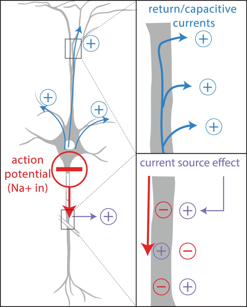
Schematic of return/capacitive currents and current source effects. Red, The extracellular spike is generated near the soma and strongly detected at the AIS. Top, Return currents (blue) describe an outflow of positive charge toward the dendrites (blue) due to the rebalance of current from the dominantly negative spike at the AIS. Bottom, The current source effect comes from the propagating action potential (red) along the axon. As the action potential traverses from the soma through the axon, negative current will flow inside the axonal membrane (negative intracellular current), while positive current exits the axonal membrane (positive extracellular current), providing a current source. The initial positive current source is indicated by the purple arrow and corresponds to the positive prepeak observed in triphasic and positive waveforms. Schematics were inspired by Heinricher (2004), Barry (2015), and Gulyás et al. (2016). All polarity signs refer to currents in the extracellular space.


